I don't know whether this is the right section to post my query. Mods kindly make the corrections if applicable. Now coming to the topic in question:
Yesterday, when I was cleaning my bare-bottom tank, I founds tens of thousands of tiny little white wormies colonizing my filter media (I'd previously cleaned my filter less than a month ago and there were no worms then). I have seen these guys on my Aquarium glass on various occasions in the past, especially when I had substrate in my tanks, but always thought that they were planaria.
This time though, I had a little bit of spare time and thought of viewing these under my school-time compound light microscope with a total magnification of 200x on a droplet mount. I was in for a surprise.
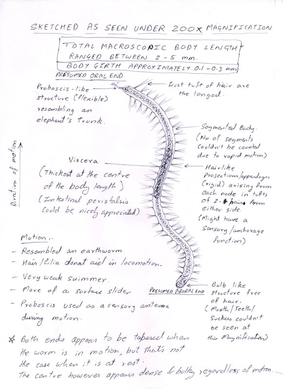
They weren't flatworms as I had expected, but were actually segmented roundworms with hairlike appendages and a highly functional flexible proboscis. The peristaltic motions of their viscera could be seen clearly, but I wasn't able to count the number of segments due to their continuous motion. I tried to overcome this by first placing a coverslip over the droplet mount (the little guy bursted) and then by absorbing the water to limit its field of motion (its body shriveled up ). But both medhods failed, ending in the death of the fragile worms.
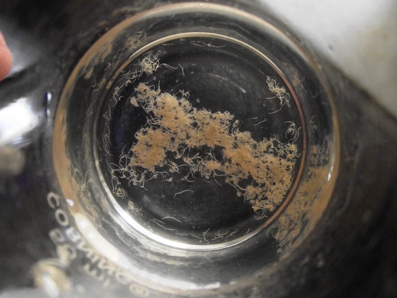
One end, which appeared to be the oral end (as motion was unidirectional) sported the proboscis, while the aboral end sported a rounded bulb like structure. The microscopic anatomy of this structure wasn't clearly visible at this magnification or at this level of activity. At the junction of every two segments, there was a tuft of rigid hairlike structures emerging from both sides. These appeared to have no active role in locomotion. These guys aren't free swimmers and only slide on the bottom and sides of the aquarium like earthworms (While stuck to the glass, their motion resembles the smooth peristaltic movements of an earthworm, but as they disengage from the glass, the movement is more of an undulant nature), but the motion is unidirectional. When sucked up and collected in a burette, these guys clumped and coiled up in the manner of tube-worms.
I tried to google them up on the internet, but was unsuccessful to find any images resembling them. I tried to capture their microscopic anatomy on camera, but was unsuccessful. Ultimately, I gave up and drew a rough sketch of the same. Inspite of being highly inaccurate (eg, the nodes/septae are more widely spaced than what they appear in the sketch), hope that it helps in their ID. I gather that they must be detritus feeding annelids, but need an actual ID, to take any measures for the safety of my fish. Can the experts give an exact id or atleast a few close guesses?
Thank you.
A Rough Sketch
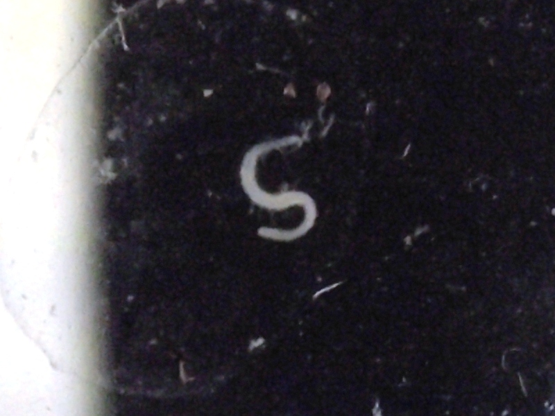
Under 20x magnification (using only the eyepiece)
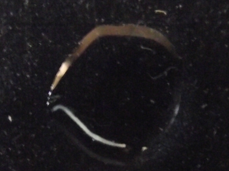
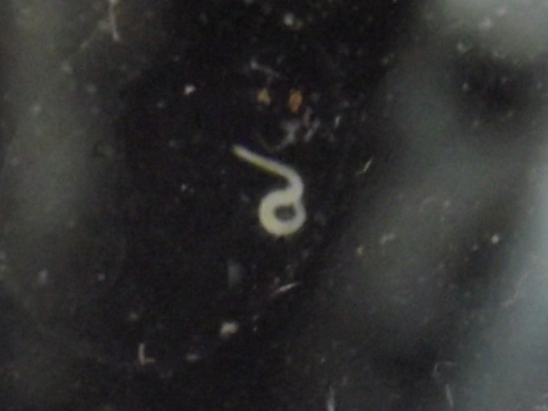
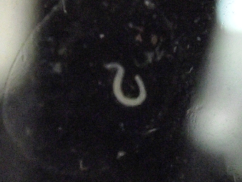
Worms in burette shot without magnification
Videos
[video=youtube;iO8eE4dw_Xg]http://www.youtube.com/watch?v=iO8eE4dw_Xg&list=UU_2sGx4ah16nyKLeXoBz1Cg&[/url] index=1&feature=plcp[/video]
Yesterday, when I was cleaning my bare-bottom tank, I founds tens of thousands of tiny little white wormies colonizing my filter media (I'd previously cleaned my filter less than a month ago and there were no worms then). I have seen these guys on my Aquarium glass on various occasions in the past, especially when I had substrate in my tanks, but always thought that they were planaria.
This time though, I had a little bit of spare time and thought of viewing these under my school-time compound light microscope with a total magnification of 200x on a droplet mount. I was in for a surprise.
They weren't flatworms as I had expected, but were actually segmented roundworms with hairlike appendages and a highly functional flexible proboscis. The peristaltic motions of their viscera could be seen clearly, but I wasn't able to count the number of segments due to their continuous motion. I tried to overcome this by first placing a coverslip over the droplet mount (the little guy bursted) and then by absorbing the water to limit its field of motion (its body shriveled up ). But both medhods failed, ending in the death of the fragile worms.
One end, which appeared to be the oral end (as motion was unidirectional) sported the proboscis, while the aboral end sported a rounded bulb like structure. The microscopic anatomy of this structure wasn't clearly visible at this magnification or at this level of activity. At the junction of every two segments, there was a tuft of rigid hairlike structures emerging from both sides. These appeared to have no active role in locomotion. These guys aren't free swimmers and only slide on the bottom and sides of the aquarium like earthworms (While stuck to the glass, their motion resembles the smooth peristaltic movements of an earthworm, but as they disengage from the glass, the movement is more of an undulant nature), but the motion is unidirectional. When sucked up and collected in a burette, these guys clumped and coiled up in the manner of tube-worms.
I tried to google them up on the internet, but was unsuccessful to find any images resembling them. I tried to capture their microscopic anatomy on camera, but was unsuccessful. Ultimately, I gave up and drew a rough sketch of the same. Inspite of being highly inaccurate (eg, the nodes/septae are more widely spaced than what they appear in the sketch), hope that it helps in their ID. I gather that they must be detritus feeding annelids, but need an actual ID, to take any measures for the safety of my fish. Can the experts give an exact id or atleast a few close guesses?
Thank you.
A Rough Sketch

Under 20x magnification (using only the eyepiece)


Worms in burette shot without magnification

Videos
[video=youtube;iO8eE4dw_Xg]http://www.youtube.com/watch?v=iO8eE4dw_Xg&list=UU_2sGx4ah16nyKLeXoBz1Cg&[/url] index=1&feature=plcp[/video]


